Disclaimer: This newsletter is for educational and informational purposes only and does not constitute medical, investment, or financial advice, nor does it establish a provider-patient relationship. Content may include forward-looking statements and discussions of investigational therapeutic candidates that are not FDA/EMA approved; their safety and efficacy remain unestablished and clinical outcomes are unpredictable. While we strive for accuracy, all information is provided as is without guarantees. As of the date of publication, the author holds no direct equity positions in the specific companies mentioned in this issue nor receives third-party compensation for this coverage. Please find a complete version of our disclaimers linked here.
Preface
The goal of this article is to unite tech and biotech communities by creating common ground and a common goal: attacking cancer. My hope is that this article will educate the tech community on the biology behind attacking cancer, while reminding the biotech community how technology was crucial in birthing the field of precision oncology. Put simply, we are better together!
Why am I writing this piece? Recently, two events have polarized tech and biotech communities in a way that, I believe, creates division rather than alliances.
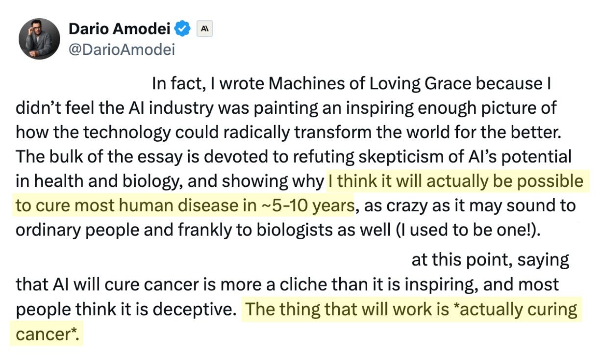
The first polarizing event is an X/tweet by Dario Amodei, Founder/CEO of artificial intelligence (AI) company Anthropic, in which he claims that AI will “cure most human disease in 5-10 years” and “the thing that [AI will achieve] is actually curing cancer”. Curing human disease and cancer are extremely noble goals that humanity should indeed strive for. However, the biotech community collectively rolled their eyes at Dario’s absurd claim that these herculean goals could be achieved with such ease and speed, let alone by a technology defined by sycophantic chatbots and slop content. But, who is more arrogant in this situation? A tech bro who believes he’s building digital God, or a biotech community that shrugs off AI as nothing but overblown hype? It’s hard to tell.
Edited and highlighted X/tweet by Dario Amodei (Founder/CEO of Anthropic) on human disease and cancer; Source: X/Twitter
The second polarizing event is the success of Moderna’s cancer vaccine in a Phase 3 clinical trial in skin cancer (stage IIB-IV melanoma), which reduced the risk of death, distant metastasis, and cancer recurrence following complete resection. The market reaction was explosive. On the day that Moderna reported the results (August 19, 2026), the company’s stock closed up 177%, making it the largest single-day jump for any S&P 500 stock of all time. Everyone cheered in unison for a victory against cancer, right? Not so fast. The tech community was quick to claim the victory as their own because they believed the drug was made using AI (it wasn’t). Dario vindicated? Cancer cured? I don’t think so. Meanwhile, the biotech community scoffed at the AI claims and grumbled at the lack of numerical data in the press release. According to New York-based analytics firm S3 Partners, hedge funds who were short Moderna stock, thinking that the company would wither away after the COVID-19 pandemic, reportedly racked up about $5.5 billion in estimated losses. Enthusiasm for Moderna’s breakthrough result came and, to quote South Park, “and it’s gone.”
Maybe we should all take a deep breath, emerge from our silos, and build a shared history of how modern medicine has been programmed to attack cancer. Perhaps the lessons of the past can guide us to a brighter future; one with less cancer. In this article, I aim to do just that by exploring landmark events in the 75-year history of precision oncology:
First Personalized Cancer Vaccine, Moderna’s intismeran autogene (investigational) ⬇️
Press ⬇️ to go down to a section and ⬆️ to go back up to the Table of Contents
Before Precision Oncology, Chemotherapy (1949)
Before we discuss precision oncology, it’s important to first remember what cancer treatment was like before those medicines came along. Prior to targeted agents, cancer treatment primarily revolved around chemotherapy, a brute force approach that uses powerful medicines to target and destroy fast-growing cells in the body. Cancer cells grow and divide much faster than normal, healthy cells. Chemotherapy drugs travel through your bloodstream to find these fast-growing cells, stopping them from reproducing or causing them to break down and die. Depending on the type and stage of cancer, doctors still use chemotherapy to this day to eradicate cancer entirely, shrink a tumor before surgery, kill any remaining cells after surgery, or control the cancer’s growth to ease symptoms.
The most-battle tested chemotherapy entered medicine from the real-life battlefield of World War II (WWII). Autopsies of World War I and II soldiers exposed to mustard gas alongside victims of a secret 1943 mustard gas bombing in Bari, Italy revealed severe suppression of bone marrow and lymphoid tissue. In August 1942, Yale pharmacologists Louis Goodman and Alfred Gilman administered synthetic nitrogen mustard (mechlorethamine) to a patient with advanced non-Hodgkin’s lymphoma (NHL), demonstrating significant tumor regression. This marked the birth of systemic cytotoxic chemotherapy. Mustargen (mechlorethamine) was formally approved by the U.S. FDA in March 1949, becoming the first FDA-approved chemotherapy.
Chemical structure of Mustargen (mechlorethamine), the first FDA-approved chemotherapy; Source: Wikimedia
A parallel effort took place on the study of antimetabolites. In 1948, Dr. Sidney Farber at Harvard demonstrated that aminopterin, a folic acid antagonist that blocks essential components for DNA synthesis, could induce temporary remissions in children with acute lymphoblastic leukemia (ALL). Farber’s research led to methotrexate, while Gertrude Elion and George Hitchings developed 6-mercaptopurine (6-MP), establishing antimetabolites as fundamental pillars of oncology.
Shortly thereafter, scientists mined the treasure trove of natural products and synthetic compounds for new chemotherapies. In the late 1950s and 1960s, researchers derived vinca alkaloids (vincristine, vinblastine) from the Madagascar periwinkle (Vinca rosea) and taxanes (paclitaxel) from the Pacific yew tree (Taxus brevifolia). Barnett Rosenberg discovered that cisplatin, a heavy-metal compound, inhibited cell division, revolutionizing testicular cancer management.
With multiple chemotherapy types at hand, researchers at the National Cancer Institute (NCI), including Emil Freireich and Emil Frei, introduced combination chemotherapy (including the four-drug cocktails abbreviated “MOPP” for Hodgkin’s disease and “VAMP” for childhood ALL). Overcoming drug resistance by hitting multiple molecular pathways simultaneously shifted cancer treatment goals from temporary palliation to potentially curative outcomes.
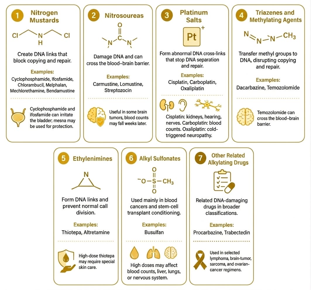
Seven classes of chemotherapy; Source: adapted from OncoDaily
But there’s a catch; a big one. Chemotherapy kills rapidly dividing cells indiscriminately, including healthy tissues. Hair follicles, bone marrow, and the gastrointestinal lining also divide quickly, so patients often experience severe side effects in these organ systems, including neutropenia, mucositis, alopecia, and severe nausea/vomiting. Repeated administration of platinum compounds, anthracyclines, or taxanes can cause cumulative cardiotoxicity, nephrotoxicity, or peripheral neuropathy. There is a glass ceiling to its efficacy as well. Multi-drug resistance (MDR) mechanisms (e.g., efflux pumps like P-glycoprotein) can cause tumors to become refractory to standard chemotherapeutics over time. Since it’s a one-size-fits-all approach, patients with the same diagnosis may receive identical regimens despite having biologically distinct tumors, leading to unpredictable response rates. To this day, chemotherapy remains a vital foundation of cancer treatment, but precision oncology agents can mitigate or, in some cases, eliminate the need for broadly toxic chemotherapy, which can dramatically improve the patient experience and outcomes.
While chemotherapy proved to be a godsend for medical treatment, one could argue that its development involved far more luck than skill. Drenching cancer in poisons that are similarly capable of extinguishing all life as we know it certainly gets the job done, but at what cost?
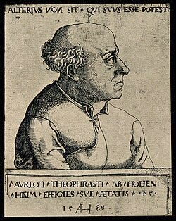
It evokes the centuries-old adage by Renaissance philosopher Paracelsus, stating, “all things are poison, and nothing is without poison; the dosage alone makes it so a thing is not a poison.” (originally written in Early New High German as “Alle Dinge sind Gift, und nichts ist ohne Gift; allein die Dosis machts, daß ein Ding kein Gift sei.”). The line appears in a controversial manuscript written by Paracelsus in 1538, titled Septem Defensiones (The Seven Defenses). Critics at the time accused Paracelsus of prescribing dangerous poisons (such as mercury and antimony). He wrote the Defenses to counter these traditional physicians, arguing that every substance (even ordinary food and water) is inherently toxic at high enough amounts, while harmful substances can be therapeutic when administered in controlled, precise micro-doses. To reiterate, the idea of micro-dosing toxins for therapeutic benefit was highly controversial in 1538, but this logic has underpinned chemotherapy since the late 20th Century. As Bob Dylan once remarked, “the times, they are a changin.”
1538 portrait of Paracelsus by Augustin Hirschvogel; Source: Wellcome Collection
Yet, scientists continued to interrogate the strangeness of cancer, hoping to make sense out of it. I’m happy to report that they found what they were looking for. The field of oncology was forever changed by a critical revelation: cancer is a disease of DNA. This revelation involved two types of genes that impact cellular replication: oncogenes (the gas pedals) and tumor suppressor genes (the brakes).
Before the mid-1970s, the leading theories suggested cancer was caused primarily by external environmental toxins, radiation, or foreign viruses. In 1976, J. Michael Bishop and Harold Varmus at UCSF discovered that the sarcoma-causing gene in the Rous Sarcoma Virus (v-src) was actually a hijacked, altered version of a gene naturally present in normal chicken cellular DNA (c-src). They proved that normal, non-cancerous cells contain precursor genes, termed proto-oncogenes, responsible for essential tasks like driving cell growth, division, and signaling. When mutated, amplified, or inappropriately expressed, these normal genes turn into hyperactive oncogenes, driving uncontrollable cell proliferation (e.g., RAS, MYC, HER2). Bishop and Varmus received the 1989 Nobel Prize in Physiology or Medicine for establishing that the seeds of cancer lie hidden in our own normal genome.
While oncogenes act as dominant gain-of-function mutations (a stuck gas pedal), scientists realized that normal cells must also possess protective mechanisms that actively stop abnormal division. In 1971, geneticist Alfred Knudson analyzed statistical patterns in pediatric retinoblastoma cases. He theorized that certain genes protect against cancer and that both alleles of these genes must be inactivated for a tumor to grow. In inherited cancer, a child receives one pre-existing “hit” (mutated allele) from a parent, requiring only one additional somatic “hit” during life to cause disease. In non-inherited cases, both copies must undergo independent, spontaneous mutations in the same cell, explaining why non-hereditary cancers develop later in life. Yet, these were mere breadcrumbs. In 1986, Thaddeus Dryja and Stephen Friend cloned the RB1 gene, confirming Knudson’s hypothesis and providing the first physical evidence of a tumor suppressor gene. Soon after, researchers recognized that p53 (encoded by TP53) acts as the ultimate tumor suppressor, often called the “guardian of the genome”, triggering DNA repair or programmed cell death (apoptosis) when damage occurs. Yet, when p53 is faulty, this cell death never occurs and tumors disseminate cells with rapidly mutating DNA throughout the body. In fact, TP53 is widely recognized as the single most common driver mutation across all human cancers. About 30% to 50% of cancers are thought to harbor somatic TP53 mutations. These mutations are found in virtually very known type of human cancer (uveal melanoma is a rare exception with nearly 0% of cases bearing TP53 mutations).
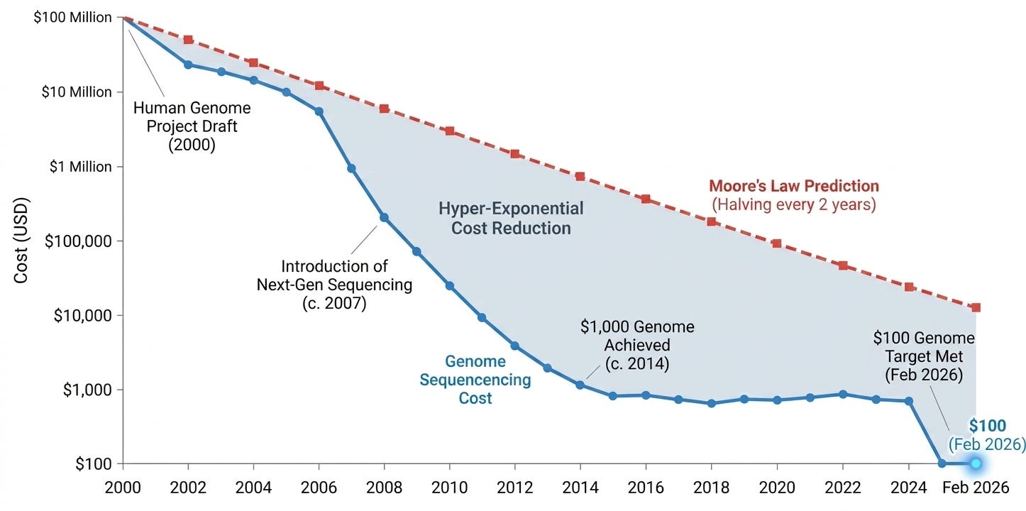
The unification of these two concepts (oncogenes & tumor suppressor genes) spurred a revolution in molecular sequencing technologies that reshaped clinical oncology. Genes like src, RB1, and TP53 were discovered by chance; by curious scientists pulling on a thread. Scientists wondered if it was possible to map the human genome and systematically identify all oncogenes & tumor suppressor genes. It began in 1990 with The Human Genome Project (HGP). The 13-year, multi-national effort to map the first complete human genome (resembling the average healthy person) established the genomic architecture needed to distinguish normal human variation from cancer-causing mutations. In the mid-2000s, high-throughput second-generation sequencing platforms replaced slow, expensive Sanger sequencing. The ability to sequence millions of DNA fragments in parallel lowered the cost of sequencing a human genome from $100 million to under $1,000, enabling widespread research and routine clinical profiling.
Launched in 2006 by the National Cancer Institute (NCI) and National Human Genome Research Institute (NHGRI), The Cancer Genome Atlas (TCGA) molecularly characterized over 20,000 primary cancer samples spanning 33 different cancer types, generating over 2.5 petabytes of multi-omic data. In that same vein, International Cancer Genome Consortium (ICGC) coordinated global efforts to catalog genomic, transcriptomic, and epigenomic alterations across thousands of tumors worldwide.
These reference datasets enabled researchers to identify specific mutational patterns left behind by carcinogens (e.g., ultraviolet radiation, tobacco smoke, or defective DNA repair pathways like BRCA1/2), providing insight into how individual tumors arise. High-resolution sequencing exposed intratumoral heterogeneity (sometimes called mosaicism), the realization that a single tumor harbors multiple sub-clones with distinct genetic profiles, explaining patients initial responded to treatment but later relapsed due to acquired resistance. Profiling expanded beyond protein-coding genes to reveal the role of promoter mutations (e.g., TERT), non-coding RNAs, and alterations in DNA methylation and histone modification in driving oncogenesis.
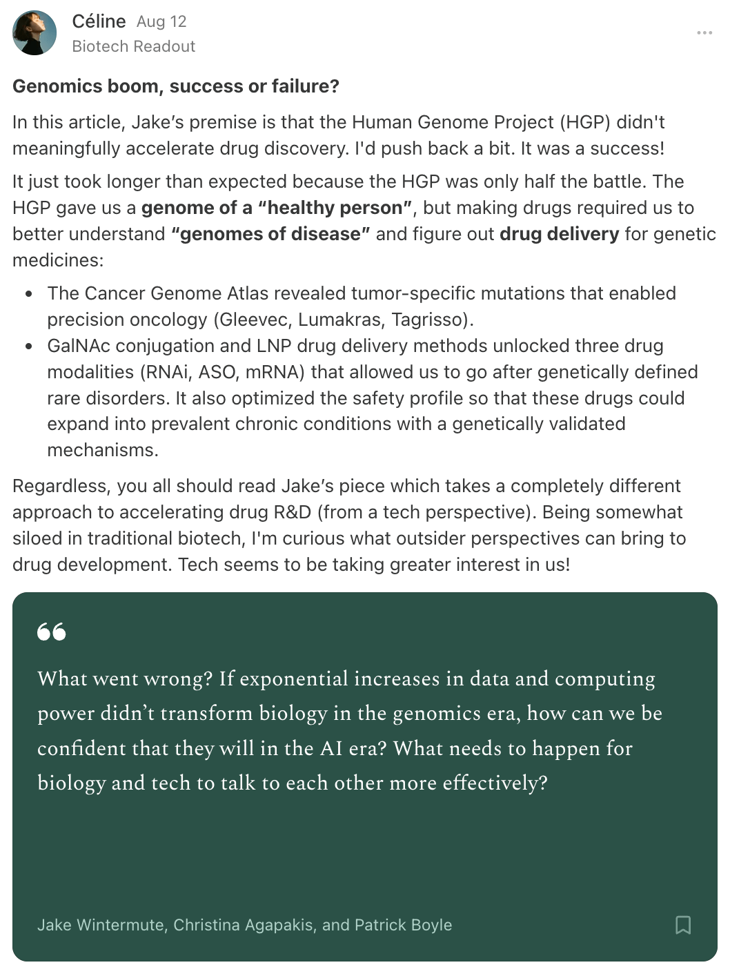
How was this deluge of answers used to sharpen our ability to program medicines to attack cancer? This is the part where I tell you that there was a third motivating factor to me writing this piece. Some claim that the genomics boom was largely fruitless and disappointing. In my Substack note below, I rebutted the often matter-of-fact claim that the Human Genome Project (HGP) didn’t meaningfully accelerate drug discovery. Well, read on, my dear readers. Read on.
My Substack note rebutting the claim that the Human Genome Project (HGP) didn’t meaningfully accelerate drug discovery
First Medicine Targeting Cell Type, Rituxan (1997)
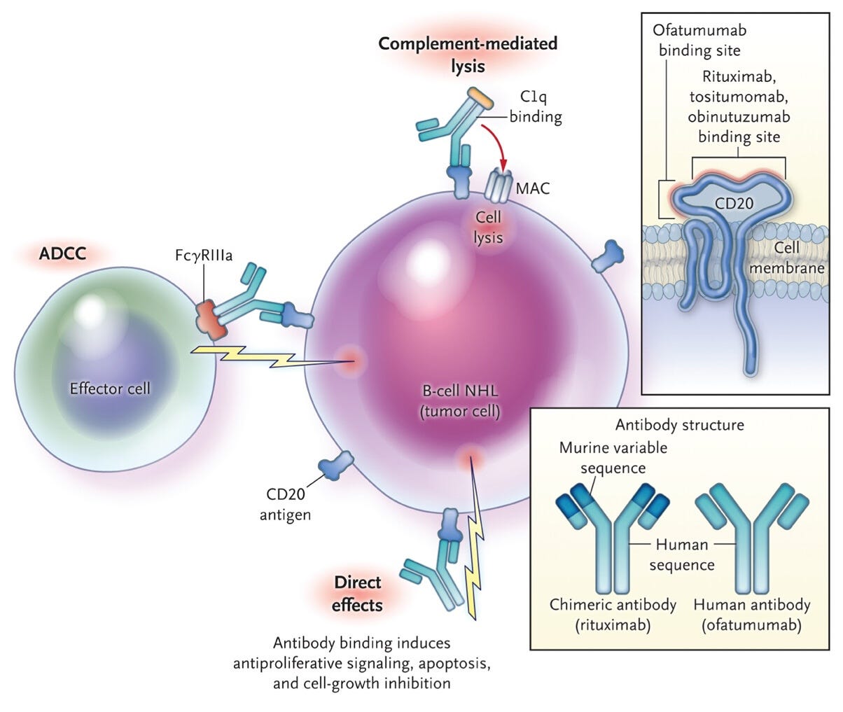
Rituxan (rituximab) was the first monoclonal antibody approved to treat cancer, marking a pivotal transition from non-specific, toxic chemotherapy to precision medicine by targeting specific cell types. The path to Rituxan began with the development of hybridoma technology in the mid-1970s by Georges J.F. Köhler and César Milstein, which allowed for the production of monoclonal antibodies (mAbs) in scaled quantities. This foundational research enabled scientists to create antibodies with predefined specificity.
In 1988, researchers identified CD20, a membrane protein found on the surface of B cells, as the ideal target. CD20 was selected because it is expressed on both malignant and mature healthy B-cells but is notably absent on immature B-cells. This meant that while the therapy would deplete cancerous B-cells, the immune system could eventually replenish healthy B-cell levels after treatment. To make the antibody viable for human use, scientists at Biogen Idec engineered Rituxan as a chimeric molecule. By fusing mouse variable regions (which provide high-affinity binding to CD20) with human constant regions, they created a therapy capable of functioning within the human body without being rapidly cleared by the immune system.
The drug’s rapid development cycle included the following milestones:
1992 - Investigational New Drug (IND) filing: An IND application was filed by Biogen Idec, only about two and a half years after the initial immunization of mice with the CD20 antigen.
1993 - Entering clinical trials: The first clinical use of rituximab occurred in a patient with lymphoma.
1997 - FDA approval: The U.S. FDA granted approval for Rituxan to treat relapsed or refractory CD20+ B-cell non-Hodgkin’s lymphoma (NHL), making it the first targeted cancer medicine approved by the agency.
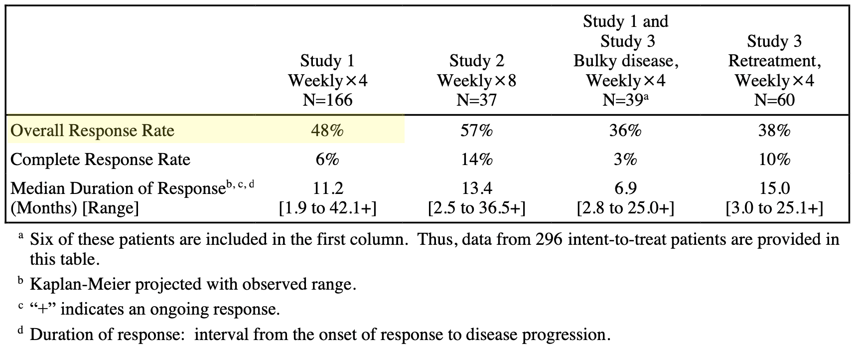
The approval of Rituxan dramatically improved the natural history of B-cell malignancies, showing an overall response rate of 48% (6% CR + 42% PR, see table below) in the pivotal, single-arm, multicenter clinical trial that supported the FDA’s decision. Following its success, monoclonal antibodies (mAbs) became a cornerstone of modern cancer treatment.
Summary of RITUXAN Efficacy Data in NHL by Schedule and Clinical Setting; Source: Rituxan label, Table 11
First Medicine Targeting Overexpressed Protein, Herceptin (1998)
Herceptin (trastuzumab) served as the first commercially successful drug to target an oncogene, a gene that, when mutated or amplified, accelerates cancer growth. The development of Herceptin began with the quest to identify specific drivers of cancer rather than relying on indiscriminate poisons, or even broad cell types like Rituxan pursued (some of which are cancer, but some of which are healthy cells).
In 1982, Robert Weinberg’s lab isolated a gene from a rat neuroblastoma called neu. By 1984, researchers at Genentech identified the human homolog of this gene, which they called HER2. Dennis Slamon, an oncologist at UCLA, collaborated with Genentech’s Axel Ullrich to test cancer cells for HER2 activity. Slamon discovered that HER2+ breast tumors were not only more aggressive and metastatic but also habitually dependent on the activity of that gene to grow. The strategy that cancer used in HER2+ breast cancer came to be known as oncogene amplification, which refers to a genetic aberration where multiple extra copies of the HER2 gene are created. This results in the excessive production (overexpression) of the HER2 protein, which drives the cancer cells to grow uncontrollably as they become habitually dependent on this signaling pathway for survival and proliferation.
In order to treat HER2+ breast cancer, oncologists needed a medicine that would erase HER2 signaling. In 1988, Genentech immunologists produced a mouse antibody that inactivated Her-2. To make it viable for human use and avoid the immune system rejecting it as a “foreign” protein, Paul Carter at Genentech “humanized” the antibody in 1990, creating trastuzumab. Its development trajectory was significantly accelerated by patient activism, which shifted the company’s internal priorities. By 1993, patients with terminal HER2+ breast cancer, led by organizations like the Breast Cancer Action project, demanded access to the drug despite it still being in clinical trials. The death of Marti Nelson, a gynecologist who had been denied access to the drug, forced a confrontation. Genentech eventually agreed to work with the National Breast Cancer Coalition (NBCC) to provide expanded access. The involvement of activists like Frances Visco (chair of the NBCC) helped move the clinical strategy from “running trials on patients” to “running trials with patients,” fundamentally changing how Phase 3 trials were recruited and managed.
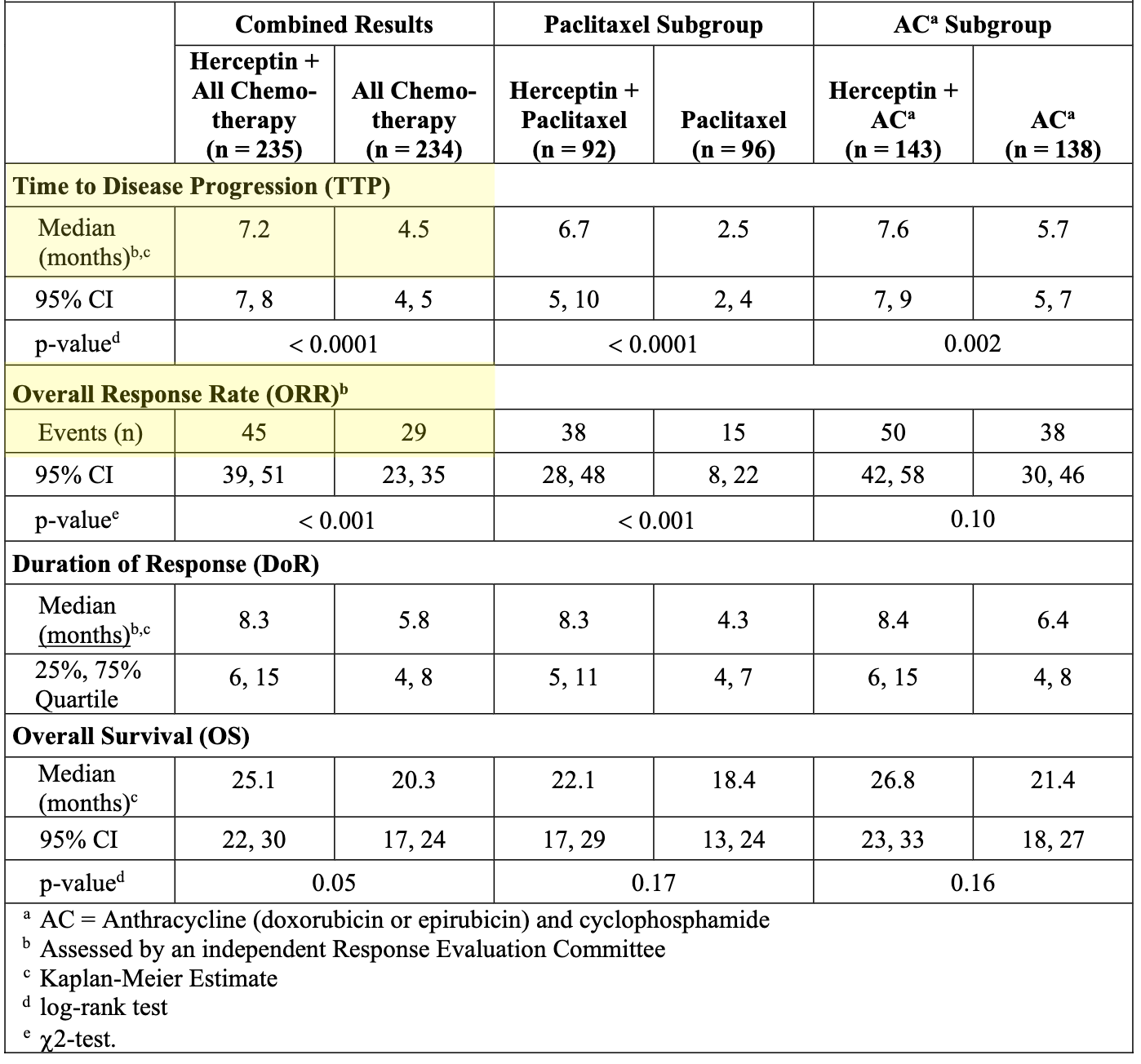
The pivotal 1998 American Society of Clinical Oncology (ASCO) meeting became the grand unveiling of the Herceptin data, a moment described by the medical community as “revolutionary”. In a multinational study of 469 women (“Trial 648”, or H0648g), the addition of Herceptin to standard cytotoxic chemotherapy increased response rates by 150% (from 29% to 45% of patients responding) and delayed cancer progression from 4.5 to 7.2 months (see table below). Subsequent studies in 2003 on early-stage, treatment-naive patients demonstrated that Herceptin combined with chemotherapy increased overall survival by approximately 33% (not shown), a magnitude of benefit unprecedented in the history of breast cancer treatment at that time. The success of Herceptin showed that targeting overexpressed proteins was a viable and powerful strategy.
Efficacy Results in First-Line Treatment for Metastatic Breast Cancer in Phase 3 H0648g trial; Source: Herceptin label, Table 11
First Medicine Targeting Cancer Mutations, Gleevec (2001)
Gleevec (imatinib) represents a landmark achievement in precision oncology, as it was the first FDA-approved cancer treatment rationally designed to target a specific cancer-causing mutation rather than broadly killing dividing cells. The development of Gleevec was driven by a precise understanding of the molecular pathogenesis of Chronic Myelogenous Leukemia (CML), a type of white blood cell cancer. This medicine targeted the so-called “Philadelphia Chromosome” and, specifically, a mutant fusion protein called BCR-ABL.
This mutation was discovered in 1959 by Peter C. Nowell, a pathologist at the University of Pennsylvania, and David A. Hungerford, a predoctoral fellow at Fox Chase Cancer Center. While studying leukemia cells under a microscope, Nowell and Hungerford observed that certain cells from patients with chronic myelogenous leukemia (CML) contained an unusually small chromosome 22. Published in 1960, their finding was significant because it provided the first evidence that cancer could be driven by a specific, identifiable genetic defect, challenging the prevailing view that cancer was unrelated to specific chromosomal changes. The abnormality was named the “Philadelphia chromosome” after the city where both researchers were based.
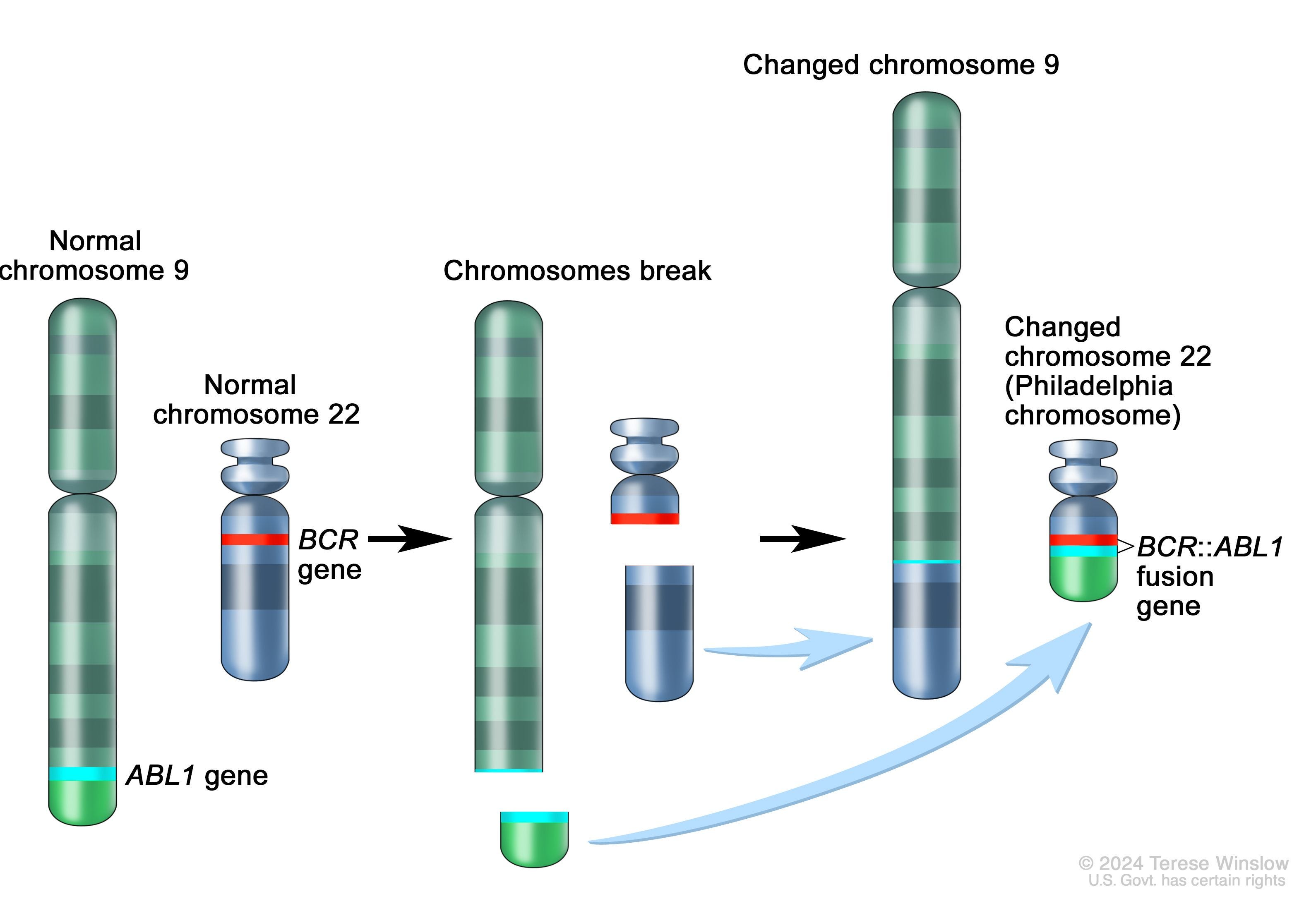
Chromosome translocation leading to short “Philadelphia chromosome” and BCR-ABL fusion protein in Chronic Myelogenous Leukemia (CML); Source: National Cancer Institute (NCI)
While the initial 1960 discovery identified the shortened chromosome 22, it wasn’t until 1973 that Janet D. Rowley discovered the full mechanism: an abnormal chromosomal translocation. In this complex biological mistake, genetic material is exchanged between chromosomes 9 and 22, specifically denoted as t(9;22)(q34;q11). This exchange fuses the ABL gene (from chromosome 9) with the BCR gene (from chromosome 22), creating the BCR-ABL fusion gene. This fusion protein acts as a constitutively active (”always on”) tyrosine kinase, which drives the uncontrolled proliferation of leukemic cells characteristic of CML.
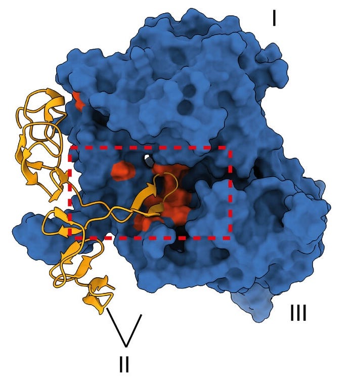
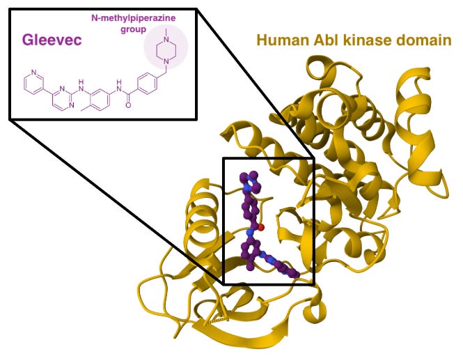
The development program for Gleevec, led by researchers at Ciba-Geigy (now Novartis) in collaboration with investigators like Dr. Brian Druker, departed from traditional drug discovery at the time (structure-based drug design is fairly commonplace today). They started by using X-ray crystallography and docking studies to optimize the molecule to bind specifically to the inactive (DFG-out) conformation of the ABL kinase domain. This unique binding mechanism contributed to its high specificity, minimizing side effects on other kinases. The N-methylpiperazine group was specifically added to the molecule to increase solubility and enhance its binding interactions within the ATP-binding pocket of the protein.
X-ray crystal structure of human Abl kinase domain (yellow) in complex with Gleevec (purple); Source: Protein Data Bank (PDB)
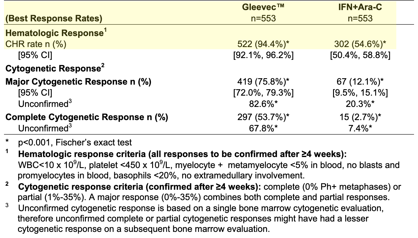
Gleevec entered clinical trials in 1998. The results seen in the initial Phase 1 and 2 trials were so unprecedented, specifically the 98% complete hematological response rate observed in chronic-phase patients, that they served as the primary clinical engine driving the drug’s rapid FDA approval, just three years after the start of Phase 1 testing. These results were confirmed by the Phase 3 IRIS trial (94.4% hematological response rate versus 54.6% standard of care [IFN+Ara-C], see table below), which compared Gleevec to the standard interferon-α plus cytarabine regimen, demonstrating Gleevec‘s clear superiority and ability to significantly improve patient survival. Following this clinical success, Gleevec received FDA approval in 2001, making it the first successful targeted therapy designed to inhibit a specific cancer mutation (BCR-ABL fusion protein).
First Medicine Unleashing the Immune System, Yervoy (2011)
The 2018 Nobel Prize in Physiology or Medicine was awarded to James P. Allison and Tasuku Honjo for their discovery of cancer therapy by inhibiting negative immune regulation. Their work established “immune checkpoint blockade” as a foundational pillar of modern cancer treatment. Allison and Honjo found that the immune system is naturally equipped with “brakes,” known as immune checkpoints, which prevent it from attacking healthy self-tissue. More specifically, James Allison identified CTLA-4 (Cytotoxic T-Lymphocyte-Associated protein 4) as a crucial negative regulator that acts as a brake on T-cell activation. Allison and his team hypothesized that tumors could be eliminated if this brake were temporarily removed. In a landmark 1996 study, they demonstrated that administering antibodies to block CTLA-4 enabled T-cells to mobilize, resulting in the rejection of tumors in mouse models of cancer.
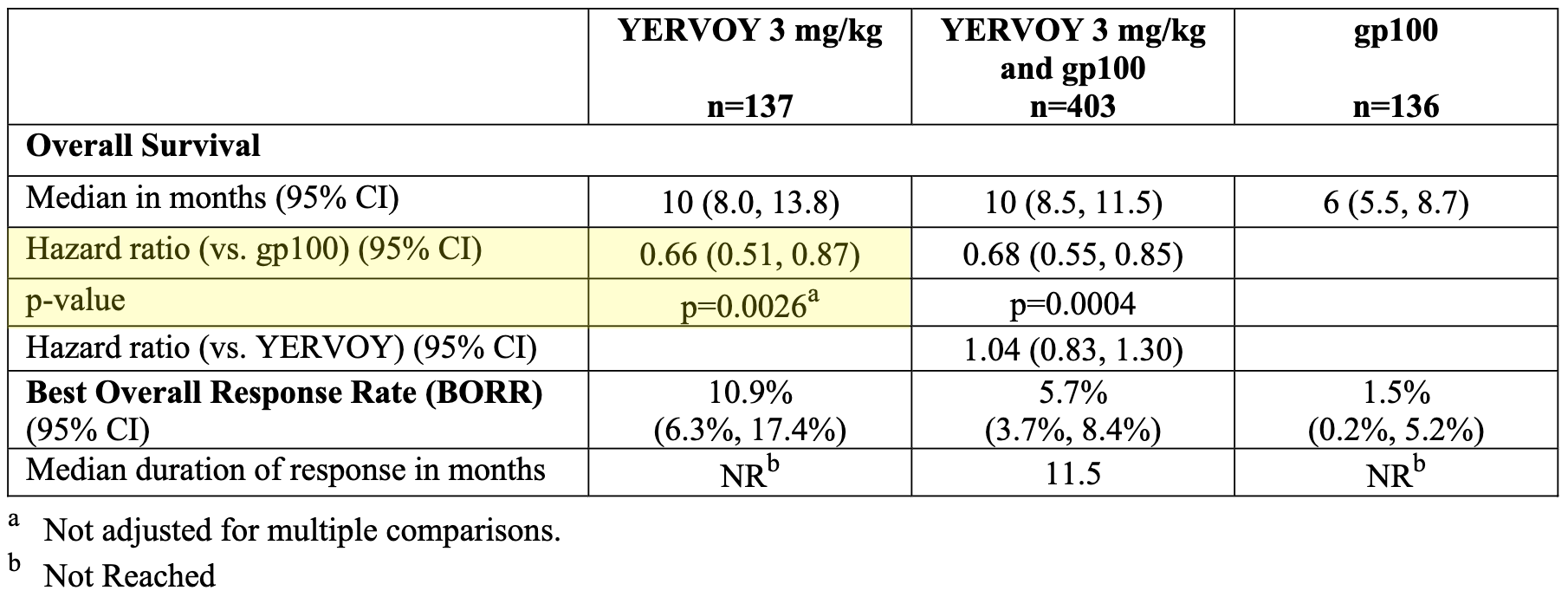
Allison’s research ultimately led to the development of Yervoy (ipilimumab), a human monoclonal antibody specifically designed to target and block CTLA-4. In 2011, Yervoy became the first immune checkpoint blockade therapy approved by the FDA for the treatment of late-stage, unresectable, or metastatic melanoma. In the trial, 676 patients with unresectable or metastatic melanoma who had already progressed on other standard therapies were randomized to receive Yervoy alone, Yervoy in combination with a vaccine, or the vaccine alone. The study demonstrated improved overall survival for patients treated with Yervoy. Specifically, the Yervoy-alone arm reduced the risk of death by 33% compared to the vaccine-only control arm (see table below).
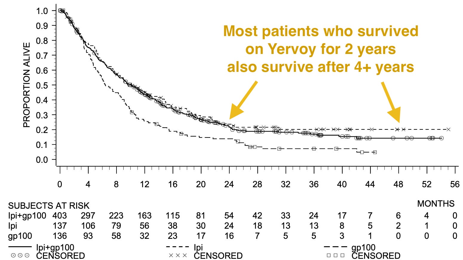
Unlike traditional chemotherapy, where responses were frequently transient, patients treated with Yervoy who responded to the drug often maintained those responses for extended periods. Reports analyzing the study results noted that as much as 70% of the objective responses observed with ipilimumab were durable. This lasting impact were believed to have contributed to the overall survival benefit seen in the trial. The estimated survival rates for patients treated with Yervoy were 46% at 1 year and 24% at 2 years, compared to 25% and 14% respectively in the comparator arm, with some patients remaining alive at 3 and 4 years (see graph below). This characteristic pattern, where the immune system is primed to continue controlling the cancer even after the course of treatment is completed, established a new treatment paradigm in oncology, distinguishing immune checkpoint blockade from the shorter-lived efficacy typically seen with cytotoxic agents.
Kaplan-Meier Curves for Overall Survival in Study MDX010-20; Source: Yervoy label, Figure 1
First Widely Used Chemo-fortified Antibodies, Adcetris (2011)
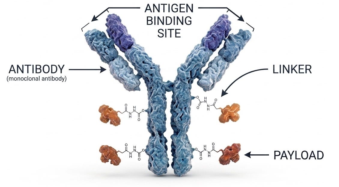
Adcetris (brentuximab vedotin), approved by the FDA in 2011, stands as the first widely successful antibody-drug conjugate (ADC), setting the standard for the modern ADC class. While Mylotarg (gemtuzumab ozogamicin) was technically the first FDA-approved ADC (in 2000), it faced significant safety concerns, leading to a voluntary withdrawal in 2010 before later being re-approved with a revised dosing schedule. For those who aren’t familiar with antibody-drug conjugates (ADCs), they consist of three component parts:
Antibody (heat-seeking missile): This is a protein designed to find and “lock onto” a specific target that is only (or mostly) found on cancer cells.
Payload (warhead): This is a highly potent chemotherapy drug. It is far too toxic to be injected into the blood on its own, as it would damage everything it touches.
Linker (safety pin): This is the chemical bond that holds the drug to the antibody. It is designed to stay stable in the bloodstream and only break once it is safely inside the cancer cell.
Diagram of antibody-drug conjugate (ADC)
Adcetris was developed to target CD30, a protein expressed on specific lymphoma cells. The drug utilizes maleimide technology to connect the cytotoxic MMAE payload to the interchain cysteine residues of an anti-CD30 monoclonal antibody (brentuximab). The VCit linker is designed to be stable in circulation, but once the ADC is internalized into a target cell, cathepsin enzymes cleave the linker, allowing for the traceless release of the payload. This “chemo-fortified antibody” approach demonstrated that targeted delivery could successfully kill malignant cells while minimizing systemic toxicity compared to conventional chemotherapy.
The FDA granted Adcetris Accelerated Approval in August 2011 based on two single-arm Phase 2 trials, using overall response rate (ORR) as a surrogate endpoint. In relapsed/Refractory Classical Hodgkin Lymphoma (cHL) pivotal Phase 2 single-arm study (n = 102) in patients who relapsed after autologous stem cell transplant (ASCT). Results showed an ORR of 73% (32% complete response, 40% partial response) with a median duration of response (DOR) of 6.7 months.
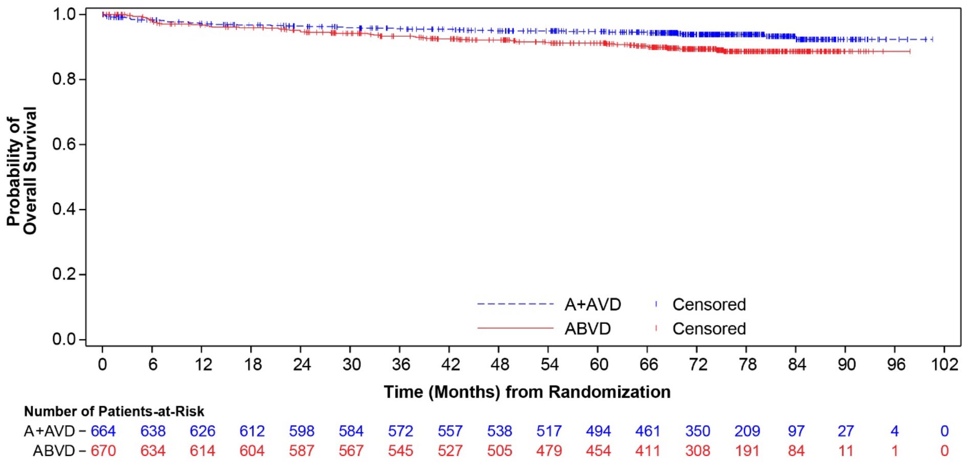
The Phase 3 trials for Adcetris were conducted post-approval to confirm clinical benefit, expand indications, and convert accelerated approvals to full regular approvals. The ECHELON-1 phase 3 trial sought to replace bleomycin with brentuximab vedotin, evaluating an A+AVD (brentuximab vedotin, doxorubicin, vinblastine, and dacarbazine) regimen against standard ABVD in over 1,300 previously untreated patients. A+AVD delivered a statistically significant improvement in progression-free survival (PFS) and overall survival (OS), representing the first frontline regimen in decades to show a survival advantage over ABVD. Long-term follow-up demonstrated a sustained 7-year OS rate of 93.5% for A+AVD compared to 88.8% for ABVD, effectively reducing the risk of death by nearly 40%.
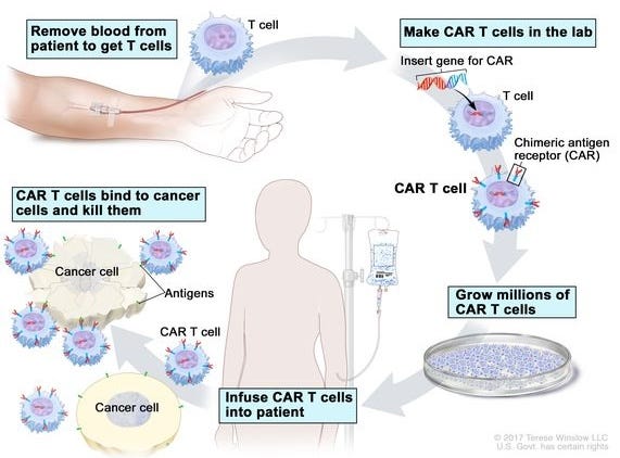
Chimeric Antigen Receptor (CAR) T-cell therapy involves genetically reprogramming a patient’s own immune system to recognize and eradicate specific cancer cells. T-cells, a type of white blood cell critical for immune defense, are isolated from the patient’s blood via leukapheresis. In a specialized lab, a disarmed viral vector inserts a synthetic gene into the T-cells’ DNA. This gene encodes the Chimeric Antigen Receptor (CAR). The newly engineered CAR-T cells are grown and multiplied into millions of doses. After brief conditioning chemotherapy to clear space in the immune system, the customized CAR-T cells are infused back into the patient.
Biotech Readout covered the development and approval of the first approved CAR-T therapy, Kymriah, in CAR-T, Part 1. To summarize:
The 2010 Landmark Trial (published in the New England Journal of Medicine in 2011) was the watershed moment [for] CAR-T therapy. While the trial was small, enrolling only three patients with advanced Chronic Lymphocytic Leukemia (CLL), the results were so explosive that they dramatically changed the trajectory of oncology. By 2010, the three patients in the trial had exhausted every available treatment, including multiple rounds of chemotherapy and monoclonal antibodies. They had several pounds of tumor mass in their bodies and were essentially in end-of-life care. After the patients were infused with their own engineered T-cells, the researchers observed a phenomena never before seen in human medicine. […] Within weeks, the pounds of tumor mass residing in the blood, bone marrow, and lymph nodes became undetectable.
When the results were published in August 2011, the headlines were staggering. The paper reported Complete Remissions (CR) in two of the three patients. Following this trial, Novartis partnered with UPenn in a landmark deal, signaling the first major Big Pharma entry into the space.
[…]
In a follow-upmore than a decade later in 2022, these same patients were still cancer-free, and the original CAR-T cells were still detectable in their blood, proving that a single infusion could potentially provide a lifetime of protection. The study focused on Doug Olson and Bill Ludwig, the two survivors from the 2010 trial. At the 10-year mark, both were still in complete remission (Bill Ludwig remained cancer-free but unfortunately passed away in January 2021 due to complications from COVID-19). Researchers found that the CAR-T cells had evolved inside the patients’ bodies. While killer CD8+ T-cells did the initial work of destroying the tumor in 2010, the long-term patrolling was carried out by a highly activated population of CD4+ CAR T-cells. Following this paper, Dr. Carl June made headlines by stating, “We can now conclude that CAR-T cells can actually cure patients with leukemia,” a rare and bold claim in oncology.
The story of Emily Whitehead is another critical milestone in the development of CAR-T therapy. If the 2011 NEJM paper demonstrated that CAR-T could work in adults, Emily demonstrated that it could work in children, and in doing so, she inadvertently taught the medical world how to manage the therapy’s most dangerous side effect: Cytokine Release Syndrome (CRS).
Emily was diagnosed with acute lymphoblastic leukemia (ALL), [a form of white blood cell cancer], in 2010 at age five. Despite 16 months of aggressive chemotherapy, her cancer relapsed twice. By early 2012, her doctors told her parents, Tom and Kari Whitehead, that she had run out of options and should enter hospice. Refusing to give up, they enrolled her in a Phase 1 clinical trial at Children’s Hospital of Philadelphia (CHOP) led by Dr. Stephan Grupp. On April 17, 2012, Emily became the first pediatric patient in the world to receive CAR-T cells.
A few days after the infusion, Emily became critically ill. This was the first time pediatricians had witnessed a full-scale Cytokine Release Syndrome (CRS). She experienced 105°F fevers, a massive drop in blood pressure, and respiratory failure. She was placed on a ventilator in the PICU and given a 1-in-1,000 chance of surviving the night. Dr. Grupp’s team ran an emergency blood test and found that her levels of Interleukin-6 (IL-6), a signaling protein for inflammation, were 1,000 times higher than normal. In one of the most famous happy accidents in medical history, Dr. Carl June’s own daughter had juvenile arthritis and took a drug called Actemra, which happens to block IL-6. Actemra had never been used for cancer-related inflammation, but with Emily near death, the team administered it as a desperate measure. Within hours, her fevers vanished. She woke up on May 2, 2012, her 7th birthday. If Emily had died, it is widely believed the FDA would have shut down the CAR-T program. Instead, her survival solidified Actemra as the gold standard of managing CRS, though the condition remains a significant and potentially life-threatening risk of CAR-T therapy.
Three weeks after Emily woke up, a bone marrow biopsy showed no signs of cancer. Emily famously stood by her father’s side when he testified before the FDA in a 2017 hearing, leading to the unanimous approval of Kymriah, the first approved CAR-T therapy. Like the adult patients in the 2022 Nature study, Emily still has patrolling CAR-T cells in her blood today. They have functioned as a permanent, living immune [defense] against her leukemia for over a decade.
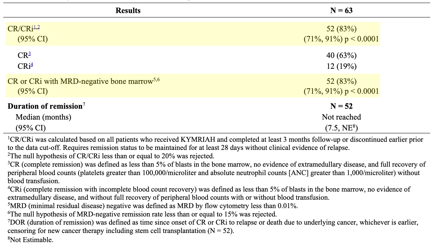
The initial FDA approval of Kymriah (tisagenlecleucel) on August 30, 2017, was granted for pediatric and young adult patients (up to 25 years old) with relapsed or refractory (r/r) B-cell precursor acute lymphoblastic leukemia (ALL). The decision was supported primary by data from the ELIANA trial, a landmark multi-center, global Phase 2 clinical trial. The trial evaluated safety and efficacy in heavily pretreated pediatric and young adult patients who had relapsed at least twice or were primary refractory to standard frontline chemotherapy. Among 63 evaluable patients followed for at least 3 months, Kymriah achieved an 83% ORR within 3 months of a single infusion. About 63% of patients achieved a Complete Remission (CR), and 19% achieved Complete Remission with incomplete blood count recovery (CRi). Among the patients who achieved remission, 99% had no detectable minimal residual disease (MRD-negative) in the bone marrow, indicating exceptionally deep molecular clearance of the cancer. The median duration of remission was not reached at the time of initial approval, demonstrating long-term persistence of the engineered cells and sustained clinical benefit.
Efficacy Results in Pediatric and Young Adult Patients with r/r B-cell ALL; Source: Kymriah label, Table 11
NOTE TO READERS: If you want to learn more about CAR-T therapy, read my three-part series on CAR-T and next-generation technologies that build on it’s success.
CAR-T, Part 1:Immunology Civil War, the discovery of T-cells, and their rebirth as medicines
CAR-T, Part 2:Beyond autologous CAR-T: allogeneic, in vivo, and T-cell engagers (TCE)
CAR-T, Part 3:How Dr. Georg Schett sparked renaissance in autoimmune disease
First Personalized Cancer Vaccine, Moderna’s intismeran autogene (investigational)
At last, we arrive at one of the medicines that motivated me to start writing this article in the first place. This is the first milestone in precision oncology for an investigational agent that is still in clinical trials, but I believe its mechanism is new enough to merit a place on this list. Allow me to separate the signal from the noise in one sentence: Moderna’s intismeran autogene is precision oncology taken to its logical extreme. Let me explain.
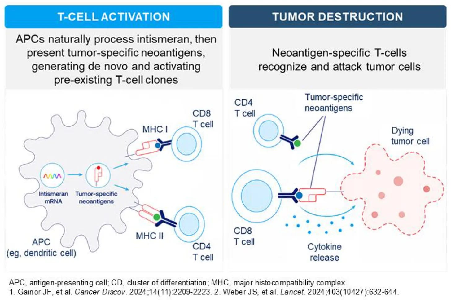
There is no other drug in the history of precision oncology that looks like this drug. Even one batch of the drug looks different than another batch! By using next-generation sequencing on resected tumor tissue, intismeran autogene encodes up to 34 patient-specific neoantigens into a single mRNA strand to prime host dendritic cells and expand cytotoxic T-cell populations against residual tumor cells. The key word there is “patient-specific”. The neoantigens that are baked into the drug cocktail for a specific patient are customized for them, based on the mutational profile of their specific tumor. Not only does this look different than any other mechanism of action, but it isn’t even one-size-fits-all like all other medicines have been since the dawn of time (x drug given for all patients with y disease). Even CAR-T therapies require insertion of the same chimeric antigen receptor (CAR) in every patient. With intismeran autogene, every patient gets a customized instantiation of the drug. Like I said, precision oncology taken to its extreme.
Let’s take a look at clinical results shown to date. We covered this recently in Weekly Review #19.
On August 19, 2026, Moderna and partner Merck announced landmark positive topline results from the Phase 3 INTerpath-001 trial evaluating intismeran autogene (mRNA-4157 / V940), an individualized neoantigen therapy (INT), in combination with Keytruda (pembrolizumab) for patients with completely resected high-risk (stage IIB–IV) cutaneous melanoma.
High-risk resected cutaneous melanoma (stages IIB–IV) is driven by oncogenic driver mutations and ultraviolet-induced DNA damage, leaving patients with a high residual risk of relapse from micrometastatic disease foci that evade immune surveillance. While current standard-of-care relies on single-agent PD-1 checkpoint inhibitors (e.g., Keytruda) or BRAF/MEK targeted therapies to reduce recurrence, intismeran autogene (mRNA-4157 / V940) offers a personalized approach as a novel synthetic neoantigen therapy. When combined with Keytruda, this regimen is designed to synergize targeted T-cell activation with checkpoint disinhibition to generate a robust cellular immune response that could prevent tumor evasion and recurrence in resected high-risk disease.
The global Phase 3 INTerpath-001 trial evaluated adjuvant treatment with intismeran autogene plus Keytruda versus Keytruda monotherapy in 1,137 adult patients with completely resected high-risk cutaneous melanoma (stages IIB–IV). At a prespecified interim analysis, the combination met its primary endpoint by delivering statistically significant and clinically meaningful improvements in Recurrence-Free Survival (RFS) compared to Keytruda alone, while also achieving statistical significance for Distant Metastasis-Free Survival (DMFS) with a manageable safety profile consistent with prior data. As the first successful Phase 3 readout for an individualized mRNA cancer therapy, these landmark results validate personalized neoantigen platforms as a viable oncology modality and establish a novel therapeutic precedent combining active, patient-specific immune priming with passive checkpoint inhibition.
Moderna and partner Merck plan to present detailed interim data, including hazard ratios and survival curves, at an upcoming medical congress. The companies will engage global regulatory authorities (such as the FDA) regarding potential supplemental biologics license application (sBLA) submissions and accelerated approval pathways. The INTerpath clinical platform is ongoing across additional Phase 2 and Phase 3 trials in non-small cell lung cancer (NSCLC), pancreatic ductal adenocarcinoma, and other solid tumors.
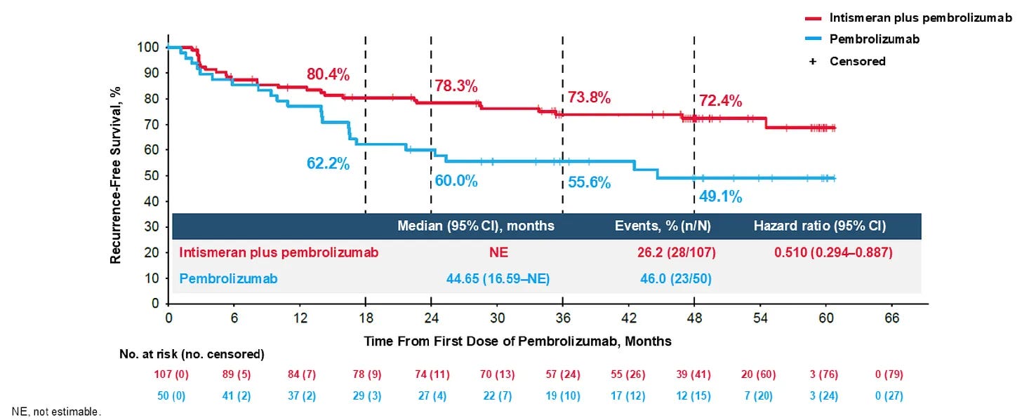
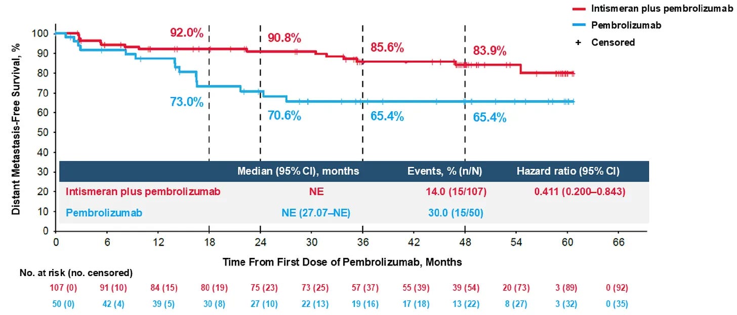
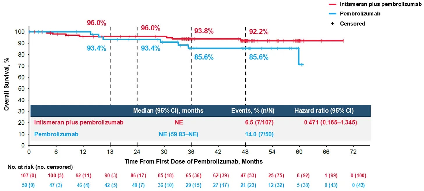
Although Moderna/Merck haven’t released Phase 3 data, the duo did present long-term 5-year follow-up data from the randomized Phase 2b KEYNOTE-942 / mRNA-4157-P201 trial in completely resected stage III/IV melanoma at the 2026 ASCO Annual Meeting (June 2026). At a median planned follow-up of 60.3 months (~5 years), adjuvant intismeran autogene plus Keytruda demonstrated a 49% reduction in the risk of recurrence or death versus Keytruda alone (HR=0.510; 95% CI: 0.294-0.887). This showed remarkable stability compared to a 3-year data cut (HR = 0.51), demonstrating that the RFS benefit did not decay over time. The combination sustained a 59% reduction in the risk of distant metastasis or death (DMFS) compared to Keytruda monotherapy (HR=0.411; 95% CI: 0.200-0.843). In an exploratory analysis, the combination showed an encouraging, clear numerical trend toward improved overall survival, with a 53% reduction in the risk of death (HR=0.471; 95% CI: 0.165-1.345), though the small sample size (n=157) limited formal statistical power. T-cell receptor (TCR) repertoire sequencing presented alongside the clinical data confirmed that the mRNA vaccine generated novel, long-lived, neoantigen-specific T-cell clones that persisted in circulation for years.
Durable Recurrence-Free Survival (RFS) benefit in resected stage III/IV melanoma in Phase 2b KEYNOTE-942 / mRNA-4157-P201 trial; Source: Moderna ASCO 2026 presentation, slide 22
Durable Distant Metastasis-Free Survival (DMFS) benefit in resected stage III/IV melanoma in Phase 2b KEYNOTE-942 / mRNA-4157-P201 trial; Source: Moderna ASCO 2026 presentation, slide 25
Overall survival in resected stage III/IV melanoma in Phase 2b KEYNOTE-942 / mRNA-4157-P201 trial shows an encouraging trend but remains immature; Source: Moderna ASCO 2026 presentation, slide 26
Precision oncology has taught us a valuable lesson: the fight against cancer is not a zero-sum game between disciplines, but a reason for tech and biotech to form a united front. The modern precision oncology armamentarium, from targeted small molecules and engineered antibodies to CAR-T therapies and personalized mRNA vaccines, was not built by biological insight or computational power alone. Instead, it was cultivated at the intersection of genomic sequencing, structural biology, and clinical execution.
As we look toward the next era of medicine, real progress will not come from hyperbolic claims that dismiss biological complexity, nor from insular skepticism that resists technological transformation. The real breakthroughs in cancer medicine will belong to those who bridge the gap: combining the speed and analytical depth of modern computing with the rigorous, empirical reality of human disease. Cancer is complex and resilient, so overcoming it will require that we bring the best tools from every field to the table.
Love biotech? Check out Biotech Readout’s full content library, or navigate directly to a segment that interests you:
Frontiers in Medicine: Exploring the frontiers of our understanding and treatments for disease.
Medical History: Recovering forgotten relics in the history of medicine.
Acquisitions: Exploring the innovation behind acquired companies.
Weekly Readout: A digest of new clinical data from the past week.
Biotech Readout is a reader-supported publication. To receive new posts and support my work, consider becoming a free or paid subscriber.
The therapeutic candidates discussed in this newsletter are currently in clinical development and have not been approved for commercial sale by the U.S. Food and Drug Administration (FDA), the European Medicines Agency (EMA), or other global regulatory authorities. Their safety and efficacy have not been established. References to pipeline products and ongoing clinical trials involve significant risks and uncertainties. Statements regarding the potential safety, potency, or efficacy of investigational drugs reflect current hypotheses and are not a guarantee of future performance or regulatory clearance. The outcome of clinical trials is inherently unpredictable, and clinical results from earlier stages may not be predictive of results in later, larger-scale trials.
No Medical Advice Disclaimer
This newsletter is for informational and educational purposes only. The content is not intended to be a substitute for professional medical advice, diagnosis, or treatment. Always seek the advice of your physician or another qualified health provider with any questions you may have regarding a medical condition. Never disregard professional medical advice or delay in seeking it because of something you have read in this publication. Investigational compounds discussed herein (including personalized cancer vaccines) remain subject to ongoing clinical evaluation and have not received regulatory approval from the FDA, EMA, or other regulatory bodies. Safety and efficacy profiles have not been established.
No Patient-Provider Relationship Disclaimer
The information provided in this newsletter is for educational and analytical purposes only. Receipt of this information, or any interaction with this content, does not create a physician-patient, pharmacist-patient, or any other professional-provider relationship between you and the authors or publishers. This newsletter should not be used as a substitute for a personal consultation with a qualified healthcare professional.
Forward-Looking Statements Disclaimer
This newsletter contains “forward-looking statements” regarding future events, including clinical trial timing, regulatory milestones, and projected market performance. These statements are based on current expectations and assumptions that are subject to significant risks and uncertainties. Actual results may differ materially from those expressed or implied. We undertake no obligation to update these statements as a result of new information or future developments.
Third-Party Links & Content Disclaimer
This newsletter contains links to third-party websites, including clinical trial registries and corporate presentations. Biotech Readout does not endorse, guarantee, or assume responsibility for the accuracy or reliability of any information offered by third-party providers.
Errors and Omissions Disclaimer
While we strive for technical accuracy, the information in this newsletter is provided on an “as is” basis with no guarantees of completeness, accuracy, or timeliness. Biotech Readout assumes no liability for any errors or omissions in the content of this publication.
Non-Endorsement Disclaimer
Any reference to specific commercial products, processes, or services by trade name, trademark, or manufacturer does not constitute or imply an endorsement or recommendation by the author. All trademarks are the property of their respective owners.
No Investment Advice Disclaimer
This newsletter is for informational purposes only and does not constitute financial, investment, or legal advice. The author is not a registered investment advisor. You should consult with a professional financial advisor before making any investment decisions. The biotechnology sector is highly volatile; past performance is not indicative of future results.
Conflict of Interest Disclaimer
The author of this newsletter maintains a position of independence. At the time of publication, the author holds no direct financial interest, equity, or options in any of the companies mentioned in this report. No compensation has been received from any third party to feature or analyze specific therapeutic candidates or corporate entities.
This is an enormous amount of work and it shows. Building the whole history as a ladder of increasing specificity, from Mustargen killing anything that divides through to Gleevec targeting one fusion protein, is what makes the last section land rather than read as hype.
Your line about intismeran being precision oncology taken to its logical extreme is the sharpest thing here, and the CAR-T comparison is what proves it. Every rung below it, CAR-T included, is one molecule or one construct given to a population. Intismeran isn't. Which makes it a different kind of object to evaluate. What INTerpath-001 is testing isn't a drug's effect, it's how reliably a pipeline (sequencing, neoantigen selection, manufacturing) produces a good product for every single patient in that arm. The hazard ratio is a statement about process consistency as much as biology.
That's probably why the missing numbers in the August release bothered people more than usual, and why the mature KEYNOTE-942 curves you pulled in do more work than the topline ever could.
Good read with good overview of the landscape. Since you ended on intismeran wanted to get your thoughts on whether you think the industry will move towards more precision and personalized therapies or do you think that more off-the-shelf therapies (ie. in vivo CAR-T) will be the standard? Or perhaps both? Just curious about your thoughts.





This is an enormous amount of work and it shows. Building the whole history as a ladder of increasing specificity, from Mustargen killing anything that divides through to Gleevec targeting one fusion protein, is what makes the last section land rather than read as hype.
Your line about intismeran being precision oncology taken to its logical extreme is the sharpest thing here, and the CAR-T comparison is what proves it. Every rung below it, CAR-T included, is one molecule or one construct given to a population. Intismeran isn't. Which makes it a different kind of object to evaluate. What INTerpath-001 is testing isn't a drug's effect, it's how reliably a pipeline (sequencing, neoantigen selection, manufacturing) produces a good product for every single patient in that arm. The hazard ratio is a statement about process consistency as much as biology.
That's probably why the missing numbers in the August release bothered people more than usual, and why the mature KEYNOTE-942 curves you pulled in do more work than the topline ever could.
Good read with good overview of the landscape. Since you ended on intismeran wanted to get your thoughts on whether you think the industry will move towards more precision and personalized therapies or do you think that more off-the-shelf therapies (ie. in vivo CAR-T) will be the standard? Or perhaps both? Just curious about your thoughts.